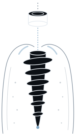
Failing bridge to fixed teeth in one day.
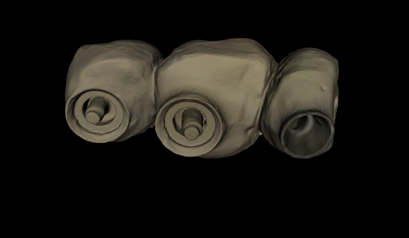
Three implants under a low sinus floor at 25, 26 and 27 — tilted to find the bone, restored on angulated multi-unit abutments, and carrying a fixed provisional before the patient left.
The case
A bridge on 25–27 was failing. Rather than replace it with another bridge on
compromised abutments, the span was converted to implants — the teeth out and the
implants in on the same day, with a fixed provisional fitted before the patient left.
The sinus, and the answer to it
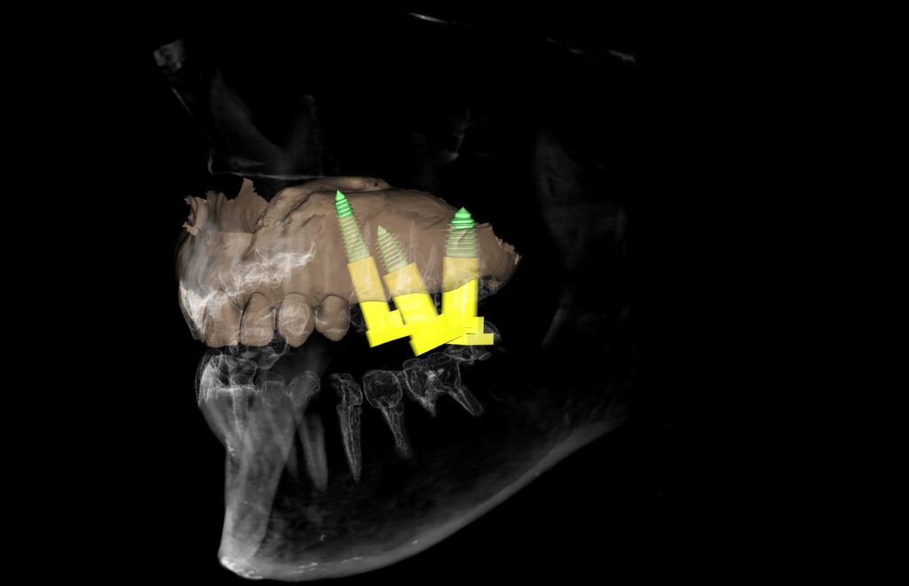
The maxillary sinus floor sits low over this quadrant. That is the constraint the whole
plan is built around, and it shows up twice.
In the fixture selection. Site 25 took a Ø 4.0 × 13 mm
implant; 26 and 27 took Ø 5.0 and Ø 6.0 at 8.5 mm. Long and narrow at the
front of the span, short and wide at the back. Where vertical bone runs out, width buys
back what length cannot.
In the angulation. The implants were tilted to find bone anterior to and
below the sinus rather than driving up into it, and restored on angulated multi-unit
abutments that bring the divergent implant axes back to a common prosthetic path. Site 25
was placed at roughly 17° off vertical.
That pairing is the point. Tilting is only useful if the prosthesis can still be made
— and the angulated abutment is what converts an implant angled for the bone into a
platform angled for the restoration. Neither half works without the other, and both were
decided on screen before anything was extracted.
The result is a posterior quadrant restored with no sinus lift, no graft and no
delay.

Why this guide
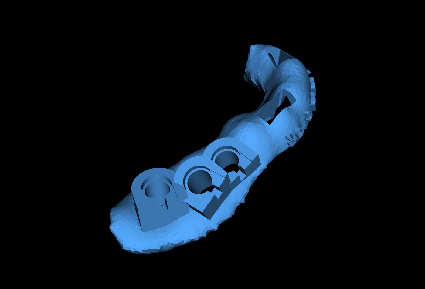
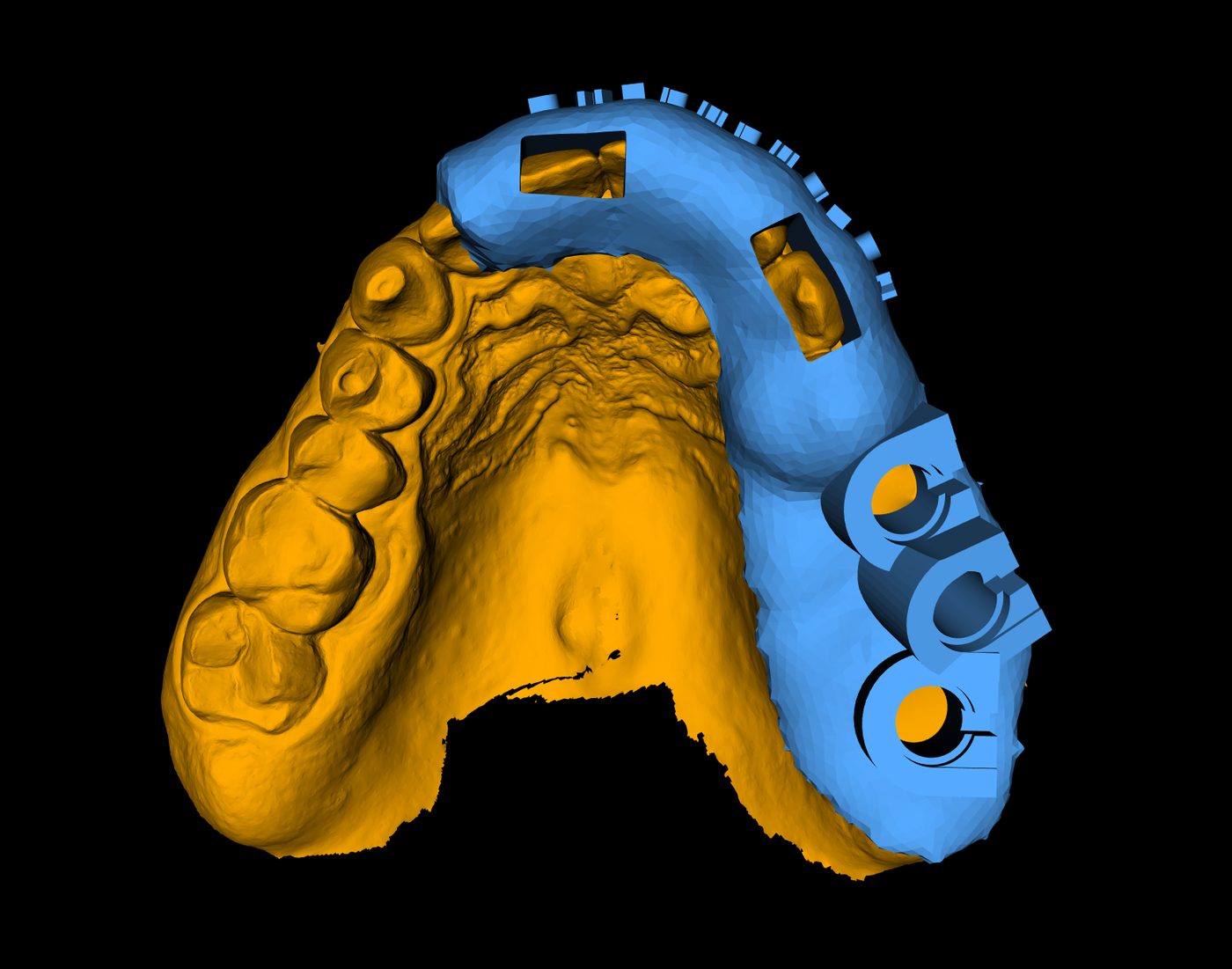
A tooth-supported template, extended onto the mucosa across the span once the teeth were
out.
The reason is the one that governs any guide over an extraction span: at the moment the
bridge abutments leave, the template loses part of what was holding it. The remaining teeth
still set the seating position and the rotational reference — they are rigid and they
cannot compress — and the tissue contact across the span stops the template flexing
between them. Teeth fix the vertical; tissue removes the bend.
The surgery was flapless: no incision, no reflection, no sutures, on a quadrant that had
just had three teeth removed.
System and software
| Site | Fixture ref | Diameter | Length |
|---|---|---|---|
| 25 | FANIHX4013 | Ø 4.0 mm | 13 mm |
| 26 | FANIHX5008C | Ø 5.0 mm | 8.5 mm |
| 27 | FALIHX6008 | Ø 6.0 mm | 8.5 mm |
Outcome
| Site | Insertion torque | ISQ |
|---|---|---|
| 25 | 45 Ncm | 82 |
| 26 | 80 Ncm | 91 |
| 27 | 93 Ncm | 89 |

In one line
A failing three-unit bridge converted to three implants in a single visit, under a sinus
that would normally mean a lift and a six-month wait — because the tilt, the fixture
and the abutment angle were all settled on screen first.