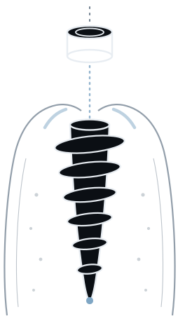
Full upper arch, eight implants, one visit.
A periodontally failing maxilla taken to a fixed provisional in a single appointment through a three-part stackable protocol — no sinus lift, no graft, no flap.
The case
A full upper arch. Severe periodontitis had left the remaining maxillary teeth mobile,
and several were already gone. The arch was not restorable tooth by tooth: what remained had
to come out, and the rehabilitation had to be planned as a single prosthetic unit from the
start.
That is the situation stackable guides exist for. Once every tooth in the arch is
leaving, the template can no longer be seated on teeth — and the moment the teeth are
extracted, the reference the plan was built on is gone. Everything downstream of that moment
has to be carried by the guide itself.
How the stack was built
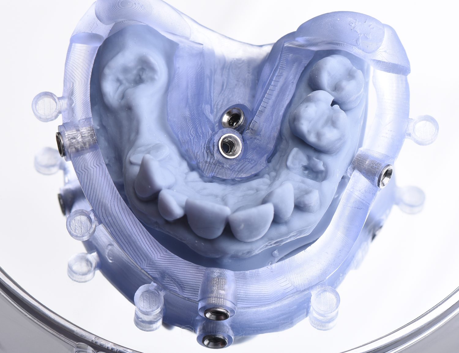
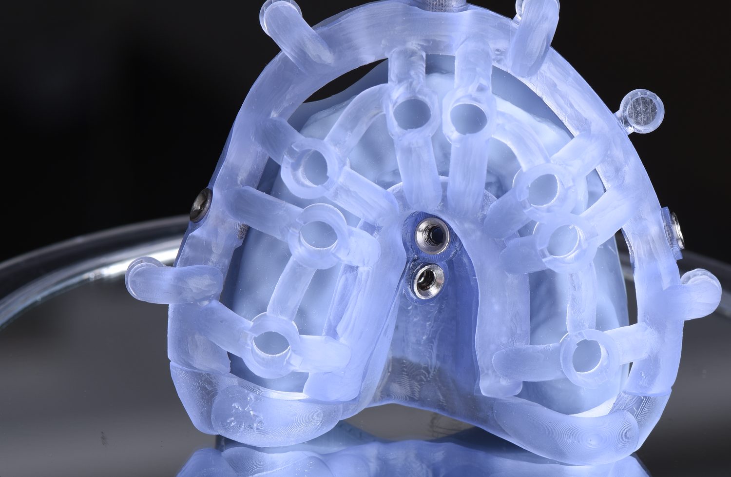
A stackable set is not one guide. It is a sequence in which each stage inherits its
position from the one before it, so that a single registration taken at the start governs
everything that follows.
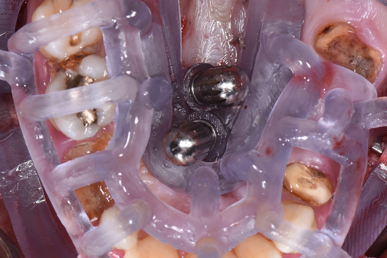
The base is pinned to bone with five fixation pins. From that moment the
arch has a coordinate system that survives the extractions: the teeth can go and the base
does not move. Every later component seats onto that same base.
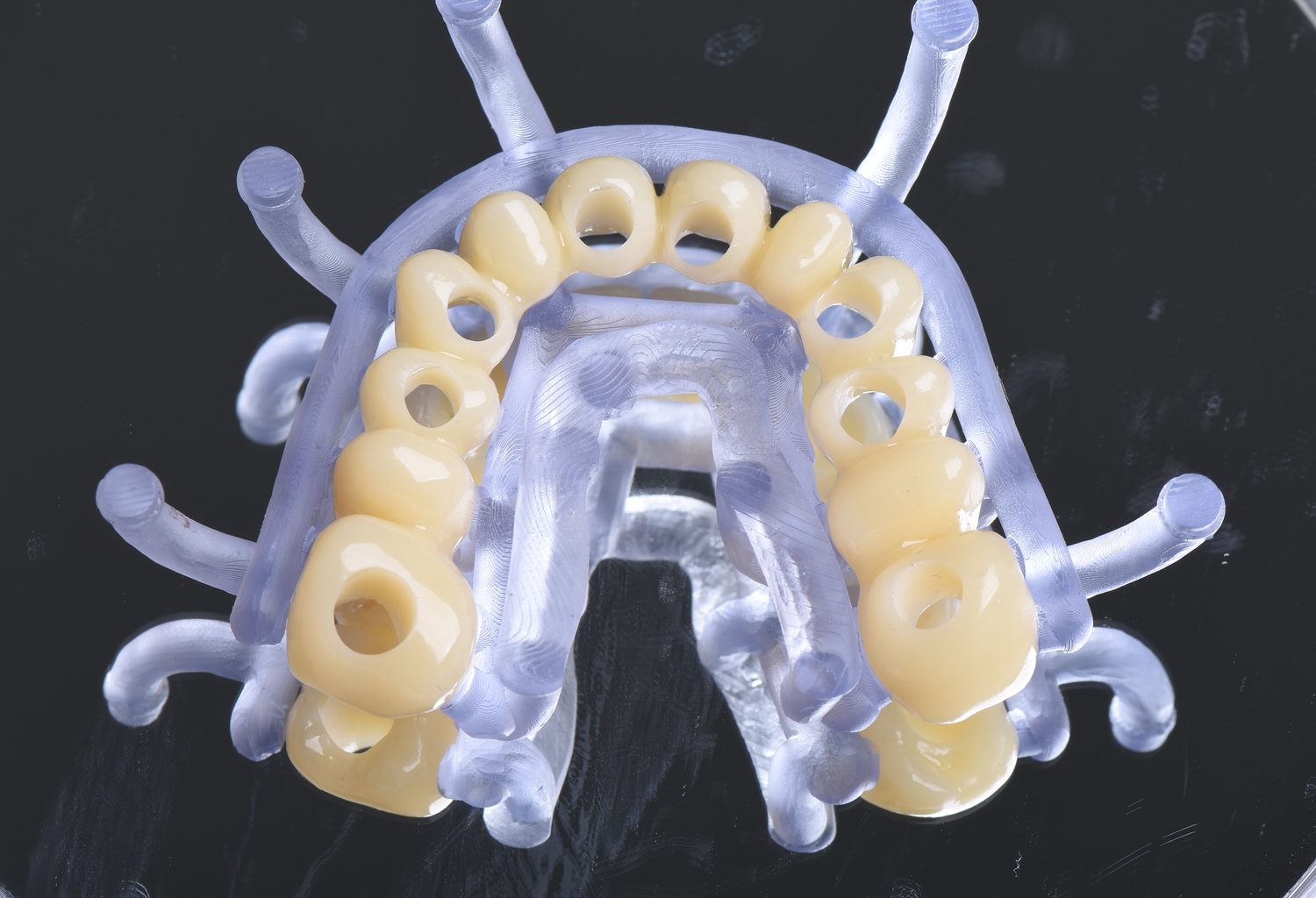
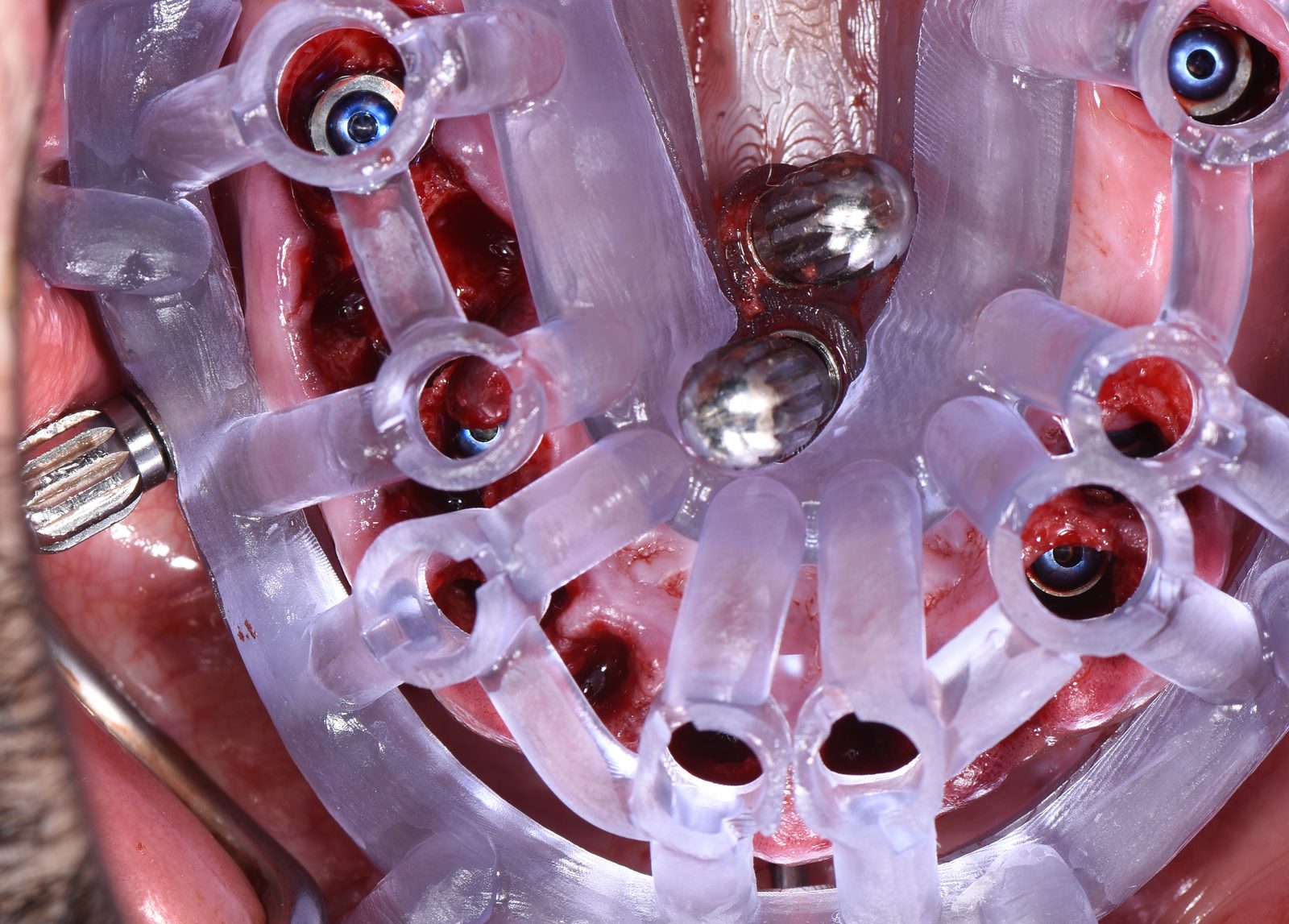
The implant guide stacks onto the base and carries the drilling protocol
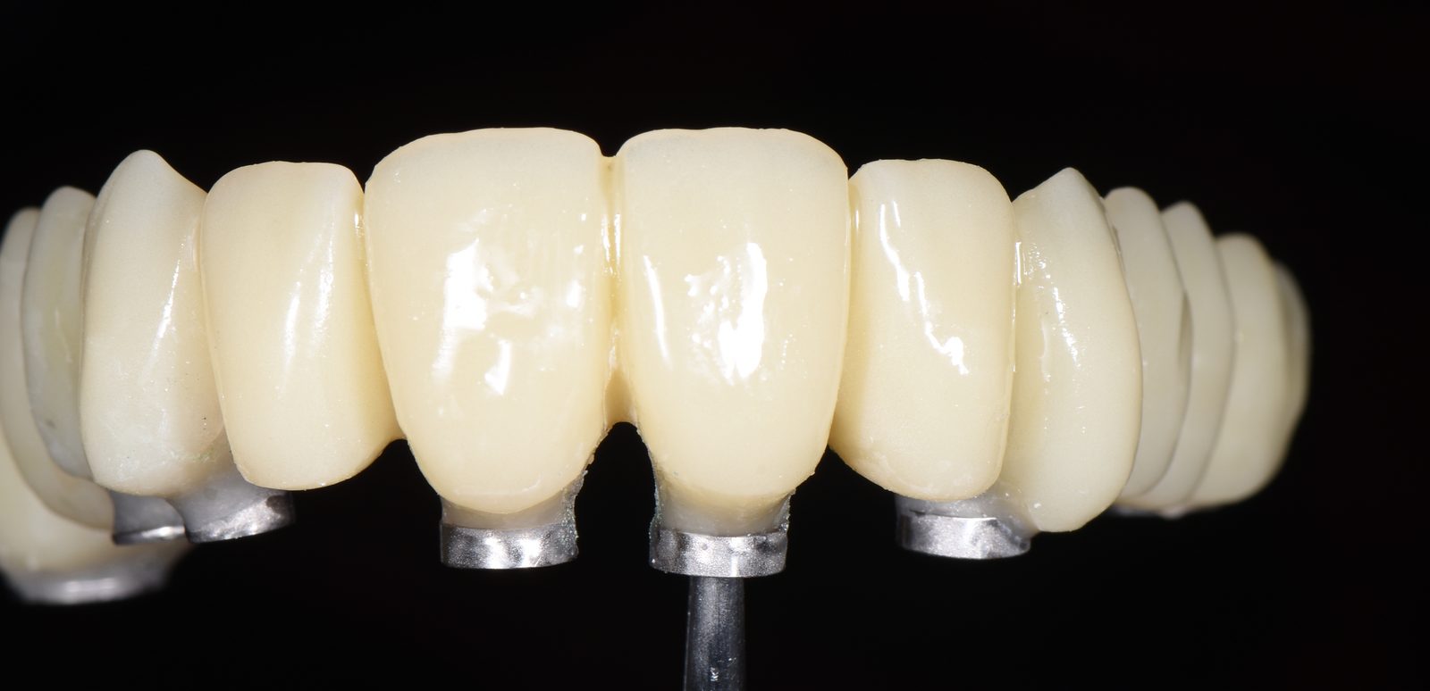
for all eight sites. The provisional guide stacks onto it in turn —
which is what fixes the occlusal position of the same-day restoration to the plan rather
than to a chairside judgement.
No bone-reduction stage was needed here. The ridge was taken as it was, which shortens
the stack to three components and removes a step at which the prosthetic plane can drift.
The point of the sequence is that nothing is re-registered mid-surgery. In a full-arch
immediate case the expensive error is not a single angulated implant — it is losing the
prosthetic reference partway through and rebuilding it by eye with the patient open in front
of you.

The distribution
Eight implants, four per side, and the two sides mirror each other exactly:
16 · 14 · 13 · 11
| 21 · 23 · 24 · 26
Nothing at the lateral incisors, nothing at the second premolars. Those become pontics.
The implants sit where the bone is worth using and where the load wants to be carried
— not at every tooth position the old dentition happened to occupy. That is the
difference between planning an arch and replacing teeth one at a time.
The sinus, and what was done about it
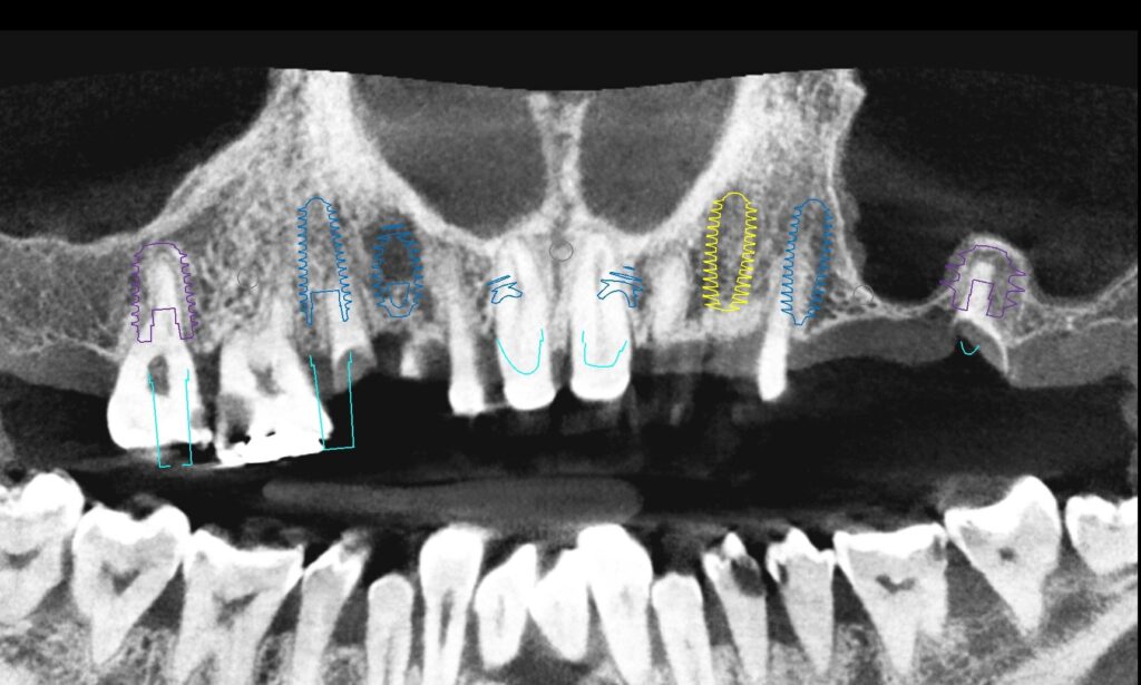
The constraint in this arch was vertical bone height under the maxillary sinuses. The
fixture list is where you can read it: site 26 took a Ø 8.0 × 7.0 mm implant and
site 16 a Ø 6.5 × 10 mm, against Ø 4.5 × 13–15 mm through the
anterior. Wide and short at both posterior ends, long and narrow in front.
At both posterior sites, 16 and 26, the implant was planned to finish at the level of the
sinus floor — approaching it rather than passing through it. Where the apex met the
floor, the intact Schneiderian membrane was tented slightly upward. The membrane was not
perforated and the floor was not broken through: a controlled transcrestal elevation carried
out at the moment of placement.
No lateral window, no biomaterial, no second surgical site, and no healing period waiting
on a graft before the arch could be loaded. The small space created beneath the elevated
membrane fills with blood clot and ossifies on its own.
This is a documented approach rather than an improvisation. Systematic-review data on
graft-free transcrestal elevation report mean endo-sinus bone gain of roughly 2–4 mm
with implant survival around 98% (Duan et al., J Periodontol 2017), and ten-year
prospective data report 3.0 ± 1.4 mm of bone gain with 100% survival (Nedir et al.,
Clin Implant Dent Relat Res 2016). Graft material is generally considered optional
where the intended elevation is under about 2 mm. Protrusion into the sinus is best kept
within 4 mm — beyond that, bone-formation efficiency falls significantly (Yu et al.,
Int J Implant Dent 2021). Planning to the floor rather than past it keeps a case at
the conservative end of that range.
It should be said plainly that this is still a sinus floor elevation, with the planning,
technique and consent that implies. Membrane perforation is reported across a wide range in
transcrestal series even in careful hands and is frequently silent; membrane thickening is
the most common radiographic finding where an implant apex protrudes into the sinus (Ragucci
et al., Int J Implant Dent 2019); and greater protrusion depth has been associated
with odontogenic sinusitis. Any maxilla planned this way is assessed for pre-existing sinus
disease first.
Which is precisely why the decision belongs in the plan and not in the surgery. Stopping
the apex at the floor rather than through it is only possible for someone who already knows,
to the tenth of a millimetre, how much bone sits beneath each posterior site — and who
can hold the drill to that depth on the day. Freehand, this anatomy is a lateral window, a
graft and a six-month delay before anything can be loaded. Planned, it is a shorter implant
of larger diameter, seated to the floor and carrying a fixed provisional the same
afternoon.

System
| Site | Fixture ref | Diameter | Length |
|---|---|---|---|
| 16 | FALIHX6510 | Ø 6.5 mm | 10 mm |
| 14 | FANIHX4513 | Ø 4.5 mm | 13 mm |
| 13 | FANIHX4515 | Ø 4.5 mm | 15 mm |
| 11 | FANIHX4513 | Ø 4.5 mm | 13 mm |
| 21 | FANIHX4513 | Ø 4.5 mm | 13 mm |
| 23 | FANIHX5013C | Ø 5.0 mm | 13 mm |
| 24 | FANIHX4513 | Ø 4.5 mm | 13 mm |
| 26 | FALIHX8007 | Ø 8.0 mm | 7 mm |
Two platforms, one case
This case was planned across both of the systems we work in, and the division was
deliberate.
R2GATE carried the prosthetic groundwork: the basic design and the
abutment selection. SMOP carried the stackable guide design — the
base, the implant guide and the provisional guide as one registered set.
That is worth spelling out rather than listing both names, because it answers the question
a reader actually has. If you run R2GATE, you would recognise the first half of this case as
your own work. If you run SMOP, the stack is where your software earns its place. Neither
tool did the whole job, and choosing which one does which part is itself part of the
planning.
The plan was shared as a link, reviewed and approved in the browser, and manufacture began
only after written approval.
The laboratory
Guide and prosthetic design by Pantelis Xydas.
Worth naming, and not only out of courtesy. In a case like this the guide design is the
surgery: the position of five fixation pins, the tolerance of three stacked components, and a
provisional made in the final design and fitted the same day are laboratory decisions, taken
before anyone picks up a handpiece. Having that work in-house rather than subcontracted is
the reason the stack and the provisional agree with each other.
Flapless
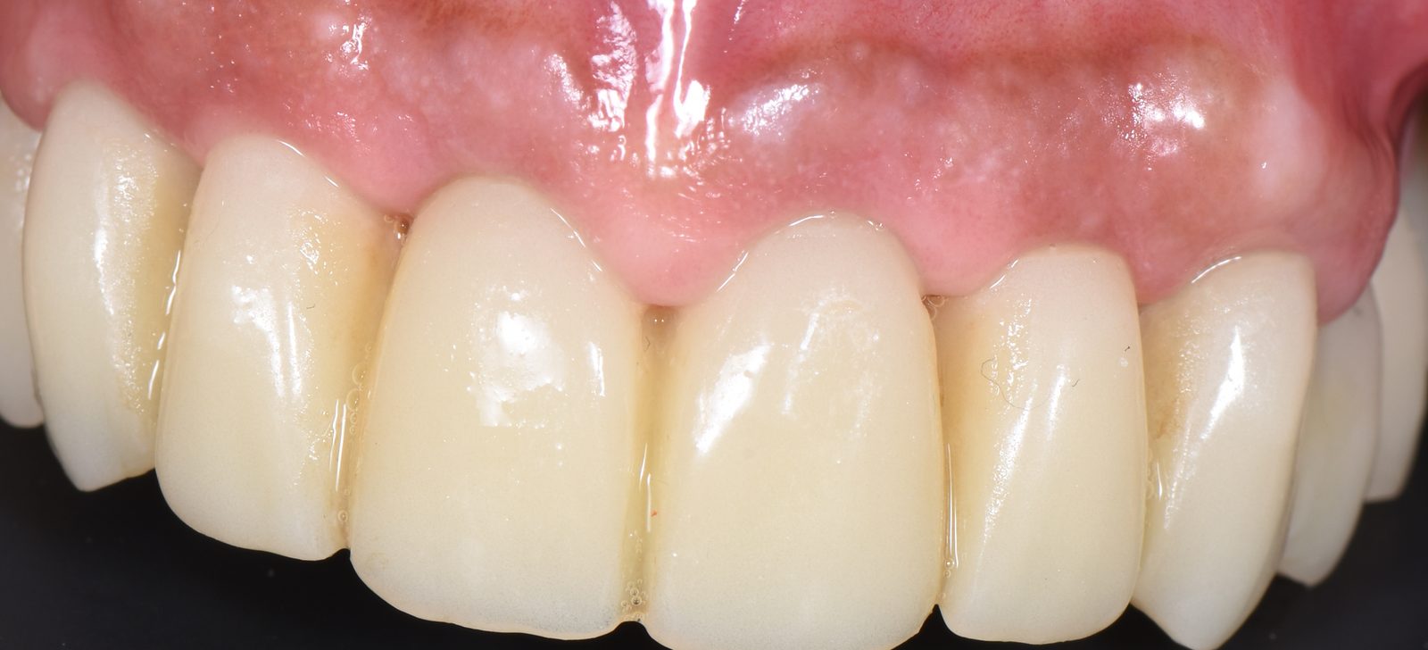
The surgery was performed flapless.
Worth stating plainly what that means here, because full-arch immediate work is usually
pictured as an open, reflected, sutured procedure. In this case the extractions, eight
osteotomies, eight implants and a fixed provisional were completed without raising a flap
— periosteum left attached, blood supply to the crestal bone undisturbed, and no
sutures in an arch that had just been cleared of periodontally involved teeth.
That is possible only because the base guide is pinned to bone. A tissue-borne template has
no answer to the compressibility of mucosa; bone fixation removes the question entirely, and
it is what lets the rest of the stack be trusted.
Outcome
| Site | Insertion torque | ISQ |
|---|---|---|
| 16 | 85 Ncm | 89 |
| 14 | 75 Ncm | 84 |
| 13 | 78 Ncm | 91 |
| 11 | 76 Ncm | 86 |
| 21 | 78 Ncm | 83 |
| 23 | 80 Ncm | 81 |
| 24 | 89 Ncm | 93 |
| 26 | 105 Ncm | 96 |
In one line
A periodontally destroyed maxilla cleared, implanted in eight sites and given fixed teeth
in one appointment — without a flap, without a graft, and without ever losing the
prosthetic reference the plan started from.